Can You Distinguish Among These Diaper Dermatoses?
Match the photographs of the rashes with the correct diagnosis.
Answers start on Next Page
Answers: 1. C; 2. E; 3. A; 4. B; 5. D; 6. F"Diaper rash" is the most common skin disease affecting infants in the western world. It is usually assumed to be caused either by irritants in the stool or urine or by a candidal infection. Although candidal diaper dermatitis and irritant contact diaper dermatitis are both extremely common, these are by no means the only causes of dermatoses in the diaper region. Here we review a number of other causes of diaper rash and provide clinical clues to help differentiate among these conditions.

1 – Irritant or Allergic Contact Diaper Dermatitis
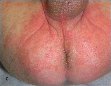
This typically presents maximally on the areas of the skin that are most in contact with the irritant or allergen (Figure C). These include the convex areas of the groin but not the hidden folds. This single finding is the most useful clue for distinguishing between contact diaper dermatitis and diaper rash caused by candidal infection-which, in contrast, favors the folds.
The most common irritants in the diaper area are stool and urine. Fragrances and harsh soaps in bathing products can also cause a dermatitis; however, when bathing products are to blame, the dermatitis is usually more generalized, extending beyond the diaper area.
The most common cause of a truly allergic diaper dermatitis is the blue dye-known as disperse blue dye-that is used in many brands of colorful diapers. The folds are almost always completely spared in infants with diaper dye contact dermatitis; also, the shape of this type of rash is a strong clue to the diagnosis. In both dye-allergy dermatitis and the more common irritant dermatitis, the edges of the rash are usually perfectly squared off.
Treatment of both irritant and allergic contact dermatitis includes removal of the offending agent and short-term use (no more than 2 weeks in most cases) of low-potency topical corticosteroid creams. Dye-free diapers are increasingly easy to find and are even sometimes less expensive than their colorful alternatives!

2 – Candidal or Monilial Diaper Dermatitis
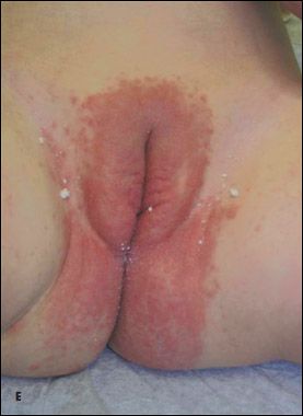
The classic clinical findings in this type of diaper rash are erythema and maceration in the inguinal creases (Figure E). The erythema is often described as "beefy red," and the maceration gives the skin the false appearance of being wet. In addition to involvement of the folds, candidal diaper dermatitis is often associated clinically with the appearance of satellite pustules emanating from the folds and forming a confluent array on the convex skin surfaces. The diagnosis is made almost solely on the basis of the clinical appearance; the history is often unhelpful, and culture is not required for confirmation.
Treatment of candidal diaper dermatitis usually consists of application of one of the topical azole antifungal creams. These agents have broader antifungal coverage than either nystatin or other over-the-counter topical antifungal therapies. The inclusion of topical corticosteroid creams in the treatment regimen is almost never necessary and is not advised. Moreover, use of combination creams that contain both antifungal agents and mid-to high-potency topical corticosteroids should be strictly avoided; such creams can be unsafe because they usually contain a higher-potency topical corticosteroid than would otherwise be prudent to use in the groin area-and because the action of the corticosteroid often counteracts the action of the antifungal in the cream.

3 – Intertrigo With Group A β-Hemolytic Streptococcal Superinfection
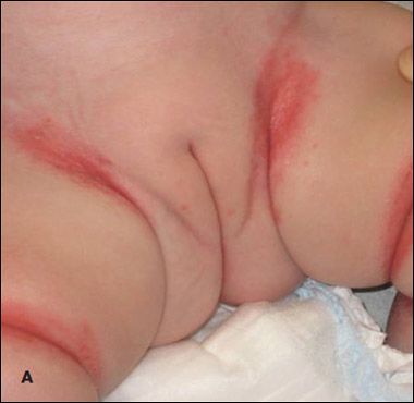
Rarely, children present with skin findings reminiscent of candidal diaper dermatitis but with extensive involvement of noninguinal areas (such as the nuchal, antecubital, and popliteal spaces) (Figure A) and an especially moist-looking appearance without satellitosis. In such cases, it is important to obtain a specimen for culture to check for group A β-hemolytic streptococci. If culture results are positive, initiate treatment with a topical antibiotic ointment, such as mupirocin ointment.
This bacterial infection usually occurs as a complication of intertrigo, a dermatitis that develops in the intertriginous areas of the skin as a result of prolonged or excessive exposure to moisture and an overgrowth of yeast. Thus, treatment also involves keeping these areas dry and applying a topical azole antifungal or wash in addition to the mupirocin to speed resolution. Unless an infant is febrile, oral antibacterial therapy is usually unnecessary.

4 – Psoriasis
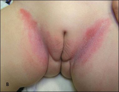
This common skin disease affects up to 2% of persons in the United States. Psoriasis is usually thought of as an adult disease; however, in 2% of affected persons, it develops before the age of 2 years. The elbows, knees, hands, feet, and scalp are the areas of the body that are most often affected. However, there is a form of psoriasis known as inverse psoriasis that affects the folds. Inverse psoriasis is seen in both adults and children; in infants it can present as a diaper rash (Figure B).
Typical clinical findings in inverse psoriasis are a diffuse, light pink erythema that starts in the groin folds; a dry appearance (as opposed to the maceration seen in candidiasis); and an absence of satellite pustules. Additional, more classic psoriatic lesions may be seen elsewhere on the body. A history of psoriasis in other family members can help confirm the diagnosis, but a family history is not always present.
Treatment of diaper psoriasis consists of intermittent use of mild topical corticosteroids, similar to those used to treat seborrheic dermatitis. If mild corticosteroids are not effective as monotherapy, topical immunomodulators may be added after a discussion of their offlabel use and black box warning is had with the family.
Seborrheic dermatitis (not pictured) is another noninfectious skin disease that commonly affects the diaper area. Although seborrheic dermatitis of the scalp, or cradle cap, is familiar to all pediatricians, seborrheic dermatitis can affect the entire body. The most common presentation of seborrheic diaper dermatitis is a diffuse diaper rash in association with classic cradle cap. The diaper rash can be differentiated from candidiasis on the basis of findings of diffuse erythema without accentuation of the folds and without satellite pustules.
Seborrheic dermatitis is thought to be at least in part a hypersensitivity reaction to an overgrowth of Malassezia yeast, which is found on the scalp and in the diaper area during the first 6 to 12 months of life. Treatment includes application of topical azole antifungal creams or washes in conjunction with intermittent use of very mild topical corticosteroids, such as hydrocortisone 1% to 2.5% cream. Again, combination creams containing both an antifungal and a mid- to high-potency corticosteroid should never be used in the groin area.
5 – Acrodermatitis Enteropathica
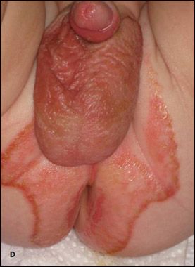
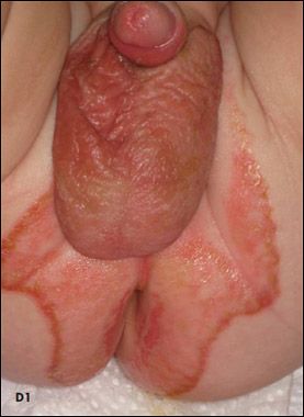
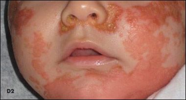
This is 1 of 2 rare and serious systemic diseases that commonly cause diaper rashes (the other is Langerhans cell histiocytosis). The rash of acrodermatitis enteropathica (AE) has been described as looking like "malignant seborrhea"-in other words, like seborrheic dermatitis, it commonly affects both the diaper area and the face; however, the typical findings of seborrheic dermatitis are accentuated and there is increased inflammation. It presents as a recalcitrant eczematous and crusted dermatitis perianally and periorally (Figures D1 and D2). The complete failure of the rash to respond to the usual topical therapy for seborrheic dermatitis also helps distinguish AE from seborrhea.
AE is associated with systemic zinc deficiency, which can result from either poor absorption of dietary zinc or malnutrition. AE typically develops when an infant is weaned from breast milk to formula; the zinc in breast milk is thought to be more bioavailable and readily absorbed than the zinc in infant formula. The exception to this clinical scenario is one in which the maternal breast milk is deficient in zinc; in this case, the deficiency-and the resultant AE-develops while the infant is still breast-feeding. AE can be diagnosed by checking the patient's zinc level or, for more rapid results, by checking the level of serum alkaline phosphatase, a zinc-dependent enzyme that can be low in cases of zinc deficiency. Treatment in most cases is with oral zinc supplementation; improvement is dramatic, usually within 1 week.

6 – Langerhans Cell Histiocytosis
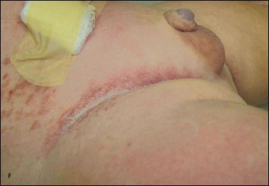
This multisystem disease can present as a severe diaper rash that is unresponsive to multiple topical therapies. Like the diaper rash of AE, Langerhans cell histiocytosis (LCH) has been described as a more malignant version of seborrheic dermatitis. Clinically, LCH appears as crusted papules with surrounding petechiae or pinpoint hemorrhage (Figure F). Lymphadenopathy may or may not be present, and if systemic progression has begun, the infant may be irritable or appear jaundiced. It is not advisable to wait for these symptoms to develop before proceeding with the systemic workup. When treating a patient with a particularly recalcitrant diaper dermatitis, it is appropriate to consider immediate referral to a pediatric dermatologist for punch biopsy to rule out cutaneous LCH. If the biopsy confirms the presence of Langerhans cells, then a full systemic workup and possible treatment with systemic therapy should be implemented by a pediatric oncologist, since diabetes insipidus, skeletal involvement, pulmonary disease, hepatic disease, or bone marrow involvement may develop in affected children.
Conclusion
Although each of the previous disease processes can present with the chief complaint of "diaper dermatitis," the astute clinician can distinguish among them on the basis of subtle differences and other clinical clues-and by careful follow-up to ensure that first-line treatment has led to the expected clinical improvement. When an infant fails to improve as expected, it is always helpful to question the original diagnosis and consider the more uncommon causes of diaper dermatitis.
Recognize & Refer: Hemangiomas in pediatrics
July 17th 2019Contemporary Pediatrics sits down exclusively with Sheila Fallon Friedlander, MD, a professor dermatology and pediatrics, to discuss the one key condition for which she believes community pediatricians should be especially aware-hemangiomas.