- Consultant for Pediatricians Vol 5 No 3
- Volume 5
- Issue 3
Photoclinic: Asymmetric Periflexural Exanthem
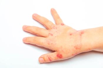
This day-old, macular, blanching, nonpruritic rash had developed in the right axilla and on the right arm and right side of the trunk of a 3 1/2-year-old boy. He was otherwise asymptomatic. Other physical examination findings were unremarkable.
This day-old, macular, blanching, nonpruritic rash had developed in the right axilla and on the right arm and right side of the trunk of a 3 1/2-year-old boy. He was otherwise asymptomatic. Other physical examination findings were unremarkable.
Sairah Chachad, MD, of Lakeland, Fla, and Deepak M. Kamat, MD, PhD, of Detroit diagnosed asymmetric periflexural exanthem--also known as unilateral laterothoracic exanthem.1 This exanthem affects children between the ages of 1 and 4 years (mean age at onset, 2 years).2 It seems to occur predominantly in females and to have seasonal variation, with peak occurrences in the spring.1,2 Only 2 familial cases have been reported.1
Most affected children are asymptomatic, although some complain of mild to moderate pruritus. A prodrome of GI or upper respiratory tract symptoms usually precedes the rash; further questioning showed this was the case in this patient. Regional lymph node enlargement and low-grade fever may occur at the time of the eruption.2,3 Despite an active search for a causative agent, the etiology remains unknown.4
The initial eruption is classically unilateral. The rash usually appears near the axilla or thorax, but it can begin in other flexures, such as the thigh, flank, or inguinal fold.2 The face, palms, and soles are spared.2 The discrete, tiny erythematous papules may become confluent patches, with a serpiginous, annular, or reticulate pattern.1 At the end of the first week, the lesions spread centrifugally to contralateral areas. The pattern is asymmetric: the side of original involvement is usually more extensively affected. After 1 to 2 weeks, the outbreak may look more scarlatiniform, morbilliform, or eczematous.2 As the lesions resolve, they leave residual dry skin.
Histologic findings are nonspecific: analysis reveals a mild to moderate mononuclear interface dermatitis with occasional necrotic and apoptotic keratinocytes.3 The dermal infiltrate is predominantly around sweat glands; there is less pronounced involvement around blood vessels and hair follicles.3 Epidermal spongiosis, as well as perivascular and periappendageal inflammation, is also present. Diagnosis is usually clinical. Establishing the diagnosis limits unnecessary testing.1
Although several conditions may mimic asymmetric periflexural exanthem, they do not share its unique unilateral distribution:
Pityriasis rosea is often preceded by a "herald patch," and the lesions are larger and more symmetric. Older children are typically affected, and the histopathologic findings differ.2
Unilateral eczematous conditions, such as allergic contact dermatitis and atopic dermatitis, cause much more itching; the lesions are weepy or crusted and respond well to topical corticosteroid therapy.2
Miliaria, which typically occurs on the face, may resemble the morbilliform lesions of asymmetric periflexural exanthem.
Roseola is transient and generalized.
Gianotti-Crosti syndrome: the lesions of this syndrome usually develop on the face and extremities--in an acral distribution.
The prognosis for children with asymmetric periflexural exanthem is excellent. The eruption usually resolves spontaneously in 3 to 6 weeks and does not recur.1 This child's rash resolved within 2 weeks. No hypopigmented or hyperpigmented areas were noted.
References:
REFERENCES:
1.
Pride HB. When a rash is more than a rash: skin signs of systemic disease.
Contemp Pediatr.
Nov 2000.
2.
Carder KR, Weston WL. Atypical viral exanthems: new rashes and variations on old themes.
Contemp Pediatr.
Feb 2002.
3.
Coustou D, Leaute-Labreze C, Bioulac-Sage P, et al. Asymmetric periflexural exanthem of childhood: a clinical, pathologic, and epidemiologic prospective study.
Arch Dermatol.
1999;135:799-803.
4.
Coustou D, Masquelier B, Lafon MD, et al. Asymmetric periflexural exanthem of childhood: microbiologic case-control study.
Pediatr Dermatol.
2000;17:169-173.
Articles in this issue
almost 20 years ago
Photoclinic: Capillary Hemangiomaalmost 20 years ago
Case in Point: Lead Poisoning in a Young Boyalmost 20 years ago
Case in Point: Thyroglossal Duct Cystalmost 20 years ago
Photoclinic: Phytophotodermatitisalmost 20 years ago
Update on Sexually Transmitted Diseases: Gonorrhea and Chlamydial Infectionsalmost 20 years ago
Photoclinic: Hirschsprung Diseasealmost 20 years ago
Winter Sports Injuries: Patterns of Injury--Preventive Measuresalmost 20 years ago
Guest Commentary: Actually, Doctor, There's This One Thing . . .almost 20 years ago
Granuloma Gluteale Infantum and Kerionalmost 20 years ago
Photo Quiz: Wart or Mimic?Newsletter
Access practical, evidence-based guidance to support better care for our youngest patients. Join our email list for the latest clinical updates.


