Photoclinic: Hirschsprung Disease
A 30-hour-old boy--born to a 36-year-old gravida 3, para 3, at full term via a spontaneous vaginal delivery--was noted to a have a mildly distended abdomen while in the newborn nursery. He had been breast-feeding every 2 to 3 hours and initially was spitting up about a quarter of the volume he had consumed. During the last 3 or 4 feedings, he had been spitting up most of the milk. There was no bilious emesis. He had not passed meconium.

A 30-hour-old boy--born to a 36-year-old gravida 3, para 3, at full term via a spontaneous vaginal delivery--was noted to a have a mildly distended abdomen while in the newborn nursery. He had been breast-feeding every 2 to 3 hours and initially was spitting up about a quarter of the volume he had consumed. During the last 3 or 4 feedings, he had been spitting up most of the milk. There was no bilious emesis. He had not passed meconium.
The newborn was awake, alert, and vigorous: weight, 3795 g (greater than 95th percentile); length, 52 cm (75th percentile); and head circumference, 35 cm (75th to 90th percentile). Head and neck findings were normal. Breath sounds were clear without murmurs. The abdomen was distended, soft, nonerythematous, with normoactive bowel sounds and no palpable masses or hepatosplenomegaly. Extremities were warm and well perfused, with good pulses. His testicles were descended bilaterally. No tufts or dimples were noted on his back. The anus was patent. On digital rectal examination, stool oozed from the anus.
Hypothyroidism, meconium ileus, Hirschsprung disease, and volvulus (with associated malrotation) were considered in the differential diagnosis.
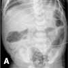
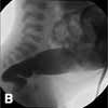
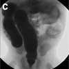
Jennifer Marin, MD, of Seattle writes that an abdominal radiograph showed dilated loops of bowel, predominantly colonic, with a relatively small rectum and wide sigmoid colon; no free air was noted (A). The infant was admitted to the neonatal ICU for further evaluation. A barium enema revealed a transition zone at the rectosigmoid junction, which suggested Hirschsprung disease (B and C). Results of a rectal biopsy confirmed the diagnosis.


Hirschsprung disease is characterized by the absence of ganglion cells in the myenteric and submucosal plexuses of the distal intestine.1 This results in absent peristalsis in the affected bowel and the development of a functional intestinal obstruction.1 The disease occurs in about 1 in 5000 live births.2
The pathogenesis and genetic basis are unclear; however, about 10% of children with the disease (especially those with longer segment disease) have a positive family history. Males are 4 times more likely to be affected than females.3 In children with Down syndrome, the incidence of Hirschsprung disease is higher; the incidence of associated congenital anomalies is about 20%.3
About 50% to 90% of cases are detected during the neonatal period.1 A distended abdomen, feeding intolerance with bilious aspirates or bilious vomiting, and delay in the passage of meconium are common findings. Typically, 95% of full-term infants pass meconium in the first 24 hours of life; fewer than 10% of children with Hirschsprung disease pass meconium during that time.1
Radiologic imaging is the first step to diagnosis. Plain films commonly show distal bowel obstruction (dilated bowel, paucity of rectal air). This test is followed by a water-soluble contrast enema; a transition zone between the normal and aganglionic bowel is pathognomonic. In about 75% of neonates with Hirschsprung disease, a transition zone is noted; therefore, its absence on contrast enema does not rule out the diagnosis.1 Rectal biopsy--the gold standard diagnostic test for Hirschsprung disease--demonstrates the absence of ganglion cells and hypertrophied nerve trunks.
Surgical repair is the definitive treatment. The goal is to remove the aganglionic bowel and bring the normally innervated bowel down to the anus, while preserving normal sphincter function.1 The most common postoperative complications include constipation, incontinence, enterocolitis, and a negative overall impact on quality of life.4
In this child, gastric decompression with a nasogastric tube was initiated. The rectum was irrigated 3 times a day and dilated twice daily until the patient underwent surgical repair. He did well postoperatively.
References:
REFERENCES:
1.
Dasgupta R, Langer JC. Hirschsprung disease.
Curr Probl Surg.
2004;41: 942-988.
2.
Swenson O. Hirschsprung's disease: a review.
Pediatrics.
2002;109:914-918.
3.
Parisi MA, Kapur RP. Genetics of Hirschsprung disease.
Curr Opin Pediatr.
2000;12:610-617.
4.
Engum SA, Grosfeld JL. Long-term results of treatment of Hirschsprung's disease.
Semin Pediatr Surg.
2004;13:273-285.