Case in Point: Lead Poisoning in a Young Boy
A 2-year-old African American boy was brought for evaluation of symptoms of an upper respiratory tract infection and intermittent abdominal pain.

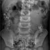
Figure 1

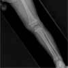
Figure 2
A 2-year-old African American boy was brought for evaluation of symptoms of an upper respiratory tract infection and intermittent abdominal pain.
He had no personal or family history of sickle cell anemia and had not been previously screened for lead poisoning.
The patient was irritable and slightly pale. Axillary temperature, 36.6°C (98°F); heart rate, 148 beats per minute; and respiration rate, 28 breaths per minute. Weight was 14.9 kg (33.1 lb). As part of a workup for apparent anemia, the blood lead level and complete blood cell count were determined. The blood lead level was 64 µg/dL, hemoglobin level was 9.6 g/dL, and mean corpuscular volume was 58.9 fL. Hemoglobin analysis ruled out sickle cell disease and glucose-6-phosphate dehydrogenase deficiency.
A radiograph of the abdomen and pelvis showed a significant amount of lead paint chips in the large intestine (Figure 1). A lower extremity radiograph revealed "lead lines," which represented abnormal calcium deposition at the distal metaphysis, not actual lead deposition (Figure 2). These lead lines suggested prolonged lead exposure.
The child was treated with polyethylene glycol to enhance elimination of the lead paint. This was followed by a 5-day course of chelation therapy with oral succimer (dimercaptosuccinic acid [DMSA]) and intravenous calcium disodium EDTA (CaNa2EDTA). He was reevaluated about 3 weeks later; the blood lead level had dropped to 33 µg/dL. His original free-erythrocyte protoporphyrin level was 1010 µg/dL.
The anemia workup revealed a low total iron level (25 µg/dL) and an elevated total iron binding capacity (555 µg/dL) and total platelet count (608 3 103/µL). It was determined that the child had hypochromic microcytic anemia caused by iron deficiency. Diet intervention and a multivitamin supplement with iron were started.
The child received chelation therapy 4 times during the following 7 months, because of reexposure and elevated rebound of lead levels.
LEAD POISONING: AN OVERVIEW
Lead poisoning continues to be a health threat to children aged 6 years and younger. The goal of the US Department of Health and Human Services is to eliminate elevated blood lead levels in children by 2010.
Lead-based paint is the most important source of lead exposure in young children. Thus, the first prophylactic measure is control of exposure to lead-contaminated soil, dust, and paint.1 Certain cities pose a high risk of lead-contaminated paint: these include Louisville and Detroit (Box).
The homes of all family members of patients with lead poisoning must be inspected by the city or county health department and deemed safe before patients return. This often requires the inspection of multiple homes (eg, grandmother's, aunt's) and may result in a search for alternative living arrangements.
Lead paint was found in this patient's home on the front porch and around the window frames. Fortunately, none of the child's older siblings had elevated blood lead levels.
DIAGNOSIS
Chronic lead poisoning is a difficult diagnosis with a wide variety of vague symptoms. Patients with lead poisoning do not typically present with symptoms of upper respiratory tract infections. This patient's symptoms were merely a fortuitous event that brought the child in for a medical evaluation. Identification of affected children often requires a heightened awareness in a community in which lead poisoning is prevalent.
Although anemia is commonly associated with lead poisoning, it is most often caused by concomitant iron deficiency--as was the case in this child.
RADIOGRAPHIC STUDIES
About 99% of children with lead poisoning who present to the Children's Hospital of Michigan have ingested lead paint. The seasonal changes in Detroit cause paint to crack and peel, which creates a potentially hazardous environment for pica in toddlers. Radiographic studies of the abdomen and pelvis must always be obtained, because chelation therapy is not started when the stomach or intestines contain paint chips. It is assumed that a concentration gradient develops when chelation therapy is begun (ie, decreased blood lead levels may cause increased lead absorption from the gut).
Radiographs of the long bones are obtained once on initial presentation. The results do not change management but can provide information about the chronicity of lead exposure. Lead lines suggest at least 6 to 8 months of lead intoxication.
MANAGEMENT
Many approaches to the ideal treatment of patients with lead poisoning are practiced, yet few data show that chelation therapy improves outcome. The CDC recommends treating children who have elevated blood lead levels of 45 to 69 µg/dL with succimer and CaNa2EDTA. Chelation with dimercaprol and CaNa2EDTA is recommended for children who have blood lead levels of 70 µg/dL or higher and for symptomatic children (those with seizures or encephalopathy).2
Few physicians manage lead poisoning with a single agent. Very few still practice the CaNa2EDTA chelation challenge test recommended in the 1972 CDC treatment guidelines.2 Although DMSA is FDA-approved only for those whose blood lead levels exceed 45 µg/dL, some physicians treat children whose blood lead levels are lower with DMSA alone, even on an outpatient basis.
Because patient compliance rates are low in Detroit, the Children's Hospital of Michigan adheres to the following protocols:
All chelation therapy is given on an inpatient basis over 5 days.
Chelation with DMSA and CaNa2EDTA is begun when blood lead levels are 40 to 69 µg/dL.
CaNa2EDTA is never given alone, because while it rapidly mobilizes lead from the bone and soft tissues, it creates a highly soluble lead-EDTA compound that may cause neurotoxicity. In addition, CaNa2EDTA is highly effective in the chelation of zinc, which results in nearly complete inactivation of d-aminolevulinic acid (ALA) dehydratase (a zinc-containing enzyme), with a consequent rise of ALA in the blood. The ALA neurotoxicity, combined with the neurotoxicity of the lead-EDTA compound, may induce severe convulsions. To prevent these acute effects, CaNa2EDTA should be given only after administration of dimercaprol or DMSA (about 4 hours later).
References:
REFERENCES:
1.
Centers for Disease Control and Prevention.
Preventing lead poisoning in young children
. Atlanta: Centers for Disease Control and Prevention; 2005.
2.
Centers for Disease Control and Prevention.
Guidelines for the management of elevated blood lead levels among young children
. Atlanta: US Department of Health and Human Services, Public Health Service, Centers for Disease Control and Prevention; 2002.