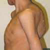
Photoclinic: Tuberculous Spondylitis
A 12-year-old boy from Pakistan presented with weakness, night sweats, anorexia, and chronic cough of 2 months' duration. He had undergone spinal surgery about 5 months before immigrating to the United States when acute paralysis, kyphosis, and a prominent midline hump (gibbus deformity) developed in his thoracic spine. The child appeared pale and weak but in no acute respiratory distress. His weight was 20.5 kg (45 lb). He had difficulty in walking without assistance. Muscle wasting was noted in the arms and legs, and he had a healing lesion on the left elbow that drained pus. Other physical examination findings were unremarkable except for a fever (temperature of 37.2°C [99°F]) and the gibbus deformity.


A 12-year-old boy from Pakistan presented with weakness, night sweats, anorexia, and chronic cough of 2 months' duration. He had undergone spinal surgery about 5 months before immigrating to the United States when acute paralysis, kyphosis, and a prominent midline hump (gibbus deformity) developed in his thoracic spine. The child appeared pale and weak but in no acute respiratory distress. His weight was 20.5 kg (45 lb). He had difficulty in walking without assistance. Muscle wasting was noted in the arms and legs, and he had a healing lesion on the left elbow that drained pus. Other physical examination findings were unremarkable except for a fever (temperature of 37.2°C [99°F]) and the gibbus deformity.
The child's father had been treated with intramuscular streptomycin for smear-positive tuberculosis (TB). A tuberculin test in the 12-year-old resulted in a 20-mm induration. A chest film demonstrated a right lower lobe infiltrate but no hilar lymphadenopathy. A bone scan showed increased activity in the T1 and T2 vertebrae. Gastric aspirates were negative for Mycobacterium tuberculosis. The lesion on the patient's elbow was attributed to TB; however, a culture of the pus did not grow M tuberculosis.
Robert Giusti, MD, and Daniel Gelfond, MD, of Long Island College Hospital in Brooklyn, NY, diagnosed tuberculous spondylitis (Pott disease).
TB is reemerging as a significant problem in areas of the United States that have a large immigrant population from countries where the disease remains endemic. Nearly 75% of TB cases occur in Pakistan, India, Bangladesh, Thailand, Indonesia, Philippines, China, Brazil, Mexico, Russia, Ethiopia, Zaire, and South Africa.1
Children contract TB from adults who have cavities in their lungs and are actively coughing up large quantities of infectious organisms. Most children with TB are not contagious because cavitary lesions rarely develop in their lungs and thus do not shed infectious organisms. Transmission is usually by inhalation of respiratory droplets, which, when dry, can remain virulent for prolonged periods. The incubation period (from infection to development of a positive tuberculin test result) is 2 to 12 weeks. The risk of tuberculous disease is highest during the 6 months after infection and remains high for 2 years.2 A diagnosis of TB in a child must be reported to the state department of public health to ensure that contact testing is performed.
The association between TB and disease of the spine that results in paralysis was first described in 1779 by Percival Pott.3 Spinal TB--the most common form of osteoarticular TB--can result from miliary (hematogenous) spread or lymphatic spread from pleural disease. Contiguous spread to the intervertebral disk produces anterior wedging of 2 or more adjacent vertebrae. In advanced disease, collapse of vertebrae results in kyphosis and a tender spinal prominence, or gibbus, as seen in this child. The upper thoracic spine is the most common site of spinal TB in children. Back pain may be the only symptom until paralysis or spinal deformity develops. Paraspinal abscesses are common; their pus can dissect and form a draining sinus or, in the case of cervical involvement, a supraclavicular mass.
The main complication of Pott disease, paraplegia, is caused by an abscess that compresses the spinal cord.4 Paraplegia can develop in the early stages of active disease or years after the initial disease has resolved.5 Patients with acute paraplegia have severe paralysis from the rapid accumulation of mechanical compression and inflammation. Weakness and paralysis of the lower extremities is a medical emergency and requires abscess drainage and vertebral stabilization. Late-onset paraplegia is best prevented by prompt diagnosis and treatment to prevent kyphosis.
Narrowing of the disk space is the earliest radiologic finding before vertebral collapse and kyphosis. A CT scan is the best modality to demonstrate the destruction of the vertebral body. Young age and good nutritional status are associated with good neurologic recovery.
This child was treated with isoniazid, rifampin, pyrazinamide, and ethambutol. Ethambutol was discontinued after 1 monthbecause it caused reversible red/green color blindness. Pyrazinamide was discontinued after 2 months. The patient completed 9 months of daily isoniazid and rifampin by directly observed therapy through the New York City Department of Health. At 1-year post-treatment, he has gained 9 kg (20 lb) and has become a vigorous and active teenager. His strength in the lower extremities is good.
References:
REFERENCES:
1.
Klein M, Iseman M. Mycobacterial infections. In: Taussig LM, Landau LI, eds.
Pediatric Respiratory Medicine.
St. Louis: Mosby; 1999:703.
2.
Pickering LK, Baker CJ, Long SS, McMillan JA, eds.
Red Book: 2006 Report of the Committee of Infectious Disease.
27th ed. Elk Grove Village, Ill: American Academy of Pediatrics; 2006:681.
3.
Pott P. The chirurgical works of Percivall Pott, F.R.S., surgeon to St. Bartholomew's Hospital, a new edition, with his last corrections. 1808.
Clin Orthop Relat Res.
2002;398:4-10.
4.
Raviglione MC, O'Brien RJ. Tuberculosis. In: Kasper DL, et al, eds.
Harrison's Principles of Internal Medicine.
16th ed. New York: McGraw-Hill; 2005:958.
5.
Jain AK. Treatment of tuberculosis of the spine with neurologic complications.
Clin Orthop Relat Res.
2002;398:75-84.